Learning to Tango

As a youngster I was introduced to most of the social graces: piano lessons – which sadly I hated – shooting, horse riding, fencing and ballroom dancing. And I just loved to tango!
Now I learn that it is, perhaps, something that more people should try.
Colleagues from the University of Turin used functional magnetic resonance imaging to study the hypothesis that simply focusing attention on walking could modify the activity of specific regions of the brain. But instead of using regular walking they taught people to tango. (The whole paper is available online as a free download.)
If you think about it for a moment, regular walking is an automatic process that you learned as a small child. It involves a series of structure in the deep – subcortical – regions of the brain and the spinal cord. The researchers got their experimental subjects to learn the dance by a combination of physical and mental practice. They were asked to perform the basic steps of the tango followed by imagining that – without moving – they were doing the steps.
The experiment showed that the training was associated with an expansion of regions of the brain involved in imagining movement. After the training the visual imagery gradually gave way to motor and kinesthetic ones.
This makes good sense: we normally begin by visualizing and then over time a motor skill like this becomes automatic. I’ve known good dancers who could flip from one dance to another without a moment’s thought.
This new finding suggests that rehabilitation of people who have lost the ability to walk should involve not just the motor act of walking, but also visualization.
But notice that the two processes – visualization and motor learning are separate. This is another nail in the coffin of the popular idea that your brain cannot tell the difference between a real and an imagined activity.
It also provides further evidence that both mental and physical activities can expand and activate regions of the brain.
And what a fun way to do it!
“The truest expression of a people is in its dance and in its music. Bodies never lie.”
–Agnes de Mille (American Choreographer, 1905-1993)
Healing the Broken Brain

I recently reviewed a fascinating book on adult neurogenesis: the creation of new neurons; something that was thought to be impossible until very recently. It is still thought to occur in certain specific regions of the brain, but even that may be changing.
The field is moving forward very rapidly and is important to every one of us, which is the reason for writing so much about the topic.
There is an exceptionally interesting article in the current issue of the journal Neuron. Researchers from Lund in Sweden have shown that cells generated from stem cells in an adult, diseased and damaged brain function as normal nerve cells. Not only do the new cells function like proper neurons, they also try to make connections with other neurons, indicating that they are trying to repair, or compensate for diseased or damaged parts of the brain.
This work was done in rats and is in its infancy, but it part of a global effort to learn more about how new neurons are formed, how they function and whether it is possible to help the brain heal itself after a disease or injury.
I am going to go out on a limb and say that it is possible. In Healing, Meaning and Purpose I described an individual with a severe neurological problem that responded to a novel treatment method using the subtle systems of the body. One of the goals of Integrated Medicine is to establish how best to use such methods in combination with conventional medicine not just to treat someone, but to initiate healing.
“A therapist doesn’t heal, he lets healing be.”
–A Course in Miracles (Book of Spiritual Principles Scribed by Dr. Helen Schucman between 1965 and 1975, and First Published in 1976)
Dreaming and Memory
As I am writing this, one of the cats is fast asleep on my desk and clearly involved in a dream that involves running and jumping. It looks as if she’s having fun.
I’ve been fascinated by dreams and dreaming ever since I read Sigmund Freud’s The Interpretation of Dreams as a teenager. For over a century, people have speculated about a link between dreams and memory, and another book that influenced me as a youngster was called Dreaming and Memory by Stanley R. Palumbo. These books were firmly rooted in a psychoanalytic framework, and I’ve always been interested in trying to reconcile psychoanalytic and neurological views of our mental life.
So I was intrigued to see an article that just came out in Nature Neuroscience.
Almost six years ago, Matthew A. Wilson at the Massachusetts Institute of Technology (MIT), in Cambridge, Massachusetts demonstrated something very interesting. Rats formed complex memories for sequences of events that they had experienced while they were awake. These memories were replayed while they slept, perhaps reflecting the animal equivalent of dreaming.
These replayed memories were detected in the hippocampus, a region of the brain that is associated with memory. But the researchers were not able to determine whether they were accompanied by the type of sensory experience that we associate with dreams-in particular, the presence of visual imagery.
In the latest research Wilson who is professor of brain and cognitive sciences at MIT’s Picower Institute for Learning and Memory, and postdoctoral associate Daoyun Ji looked at what happens in rats’ brains when they dream about the mazes they ran while they were awake. They recorded brain activity simultaneously in the hippocampus and the visual cortex, and demonstrated that replayed memories did, in fact, contain the visual images that were present during the running experience. Neurons that are activated when the animal experiences an event while awake are reactivated during sleep.
There is another piece to this research: the regions of the cerebral cortex that processes input from the senses and the hippocampus communicate with each other during sleep, leading us to speculate that this process reinforces and consolidates memories.
As the authors write, "These results imply simultaneous reactivation of coherent memory traces in the cortex and hippocampus during sleep that may contribute to or reflect the result of the memory consolidation process."
This is of great practical importance: it strongly supports the idea that adequate sleep is necessary to consolidate memory.
When I was a child my parents taught me to read through all my schoolwork just before I went to bed, even if I was tired. Work in psychology has shown that this is often the best way of memorizing factual material, and this new research shows us why.
“Waking is long and a dream short; other than this there is no difference. Just as waking happenings seem real while awake, so do those in a dream while dreaming. In dream the mind takes on another body. In both waking and dream states thoughts, names and forms occur simultaneously.”
–Ramana Maharshi (Indian Hindu Mystic and Spiritual Teacher, 1879-1950)
“The light of consciousness passes through the film of memory and throws pictures on your brain. Because of the deficient and disordered state of your brain, what you perceive is distorted and colored by feelings of like and dislike. Make your thinking orderly and free from emotional overtones, and you will see people and things as they are, with clarity and charity.”
— Sri Nisargadatta Maharaj (Indian Spiritual Teacher and Exponent of Jnana Yoga and Advaita Doctrine, 1897-1981)
Psilocybin and Mystical Experience

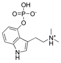
The Psilocybin Molecule
We have discussed mystical experiences a couple of times recently. They are important not only because of what they may teach us about altered states of consciousness, but because they may contain genuine revelations about the nature of reality and they are invariably profoundly meaningful to the person having them.
Earlier this year there was a paper that seemed to come in under some people’s radar, though several heavyweights in the world of psychopharmacology spoke approvingly of the research.
The paper was entitled, “Psilocybin can occasion mystical-type experiences having substantial and sustained personal meaning and spiritual significance.” There are also some superb commentaries on the original paper, all of which are available for free download if you click on the links above.
So what got everyone so excited?
Psilocybin is a psychedelic alkaloid that has been used for religious purposes for centuries. The researchers conducted a double-blind study on the acute and longer-term psychological effects of a high dose of psilocybin. What was particularly important was that the 36 experimental subjects had no previous experience of hallucinogens but who were regularly participating in religious or spiritual activities.
It is also important that the experiments were performed in comfortable, supportive surroundings. The last thing that anyone wanted was for people to have a “Bad trip” and to be left without care and support.
They were given psilocybin and methylphenidate (Ritalin) in separate sessions, the methylphenidate sessions serving as a control and active placebo; the tests were double-blind, with neither the subject nor the administrator knowing which drug was being administered. The degree of mystical experience was measured using a questionnaire on mystical experience developed by Ralph W Hood.
61% of subjects reported a “complete mystical experience” after their psilocybin session, while only 13% reported such an outcome after their experience with methylphenidate. Two months after taking psilocybin, 79% of the participants reported moderately to greatly increased life satisfaction and sense of well-being.
About 36% of participants also had a strong to extreme “experience of fear” or dysphoria (eg, a “bad trip”) at some point during the psilocybin session (which was not reported by any subject during the methylphenidate session), with about one-third of these (13% of the total) reporting that this dysphoria dominated the entire session. These negative effects were reported to be easily managed by the researchers and did not have a lasting negative effect on the subject’s sense of well-being.
The observation that psilocybin reliably elicits a transcendent, mystical state tells us that investigations of these drugs may help us understand molecular alterations in the brain that underlie mystical religious experiences.
But the key point to be made is that finding a biochemical basis for mystical experiences does nothing to belittle them. The biochemical and neurological approaches have nothing to say about the personal meaning and the cultural and social components of the experience. To use Ken Wilber’s terminology, this research is only addressing the Upper Right Hand Quadrant.
Important work to be sure, but it would be a mistake to try to reduce mystical experiences to 5HT2A,C receptors alone.
“For what is Mysticism? It is not the attempt to draw near to God, not by rites or ceremonies, but by inward disposition? Is it not merely a hard word for ‘The Kingdom of Heaven is within’? Heaven is neither a place nor a time.”
Florence Nightingale (English Pioneer of Nursing Known as the “Lady with the Lamp,” 1820-1910)
“The mystical life is the centre of all that I do and all that I think and all that I write. …I have always considered myself a voice of what I believe to be a greater renaissance – the revolt of the soul against the intellect.”
— William Butler Yeats (Irish Poet, Dramatist, Writer and, in 1923, Winner of the Nobel Prize in Literature, 1865-1939)
Head Size and Intelligence

In August we discussed some of the new data showing an association between the hormone insulin-like growth factor 1 (IGF-1) and a child’s IQ.
Now new research from the University of Southampton in England has found an association between skull size and intelligence. It seems that the brain volume a child achieves by the age of 1 year helps to determine his or her later intelligence.
The study of 633 newborns born at term has found that those with larger heads scored better on intelligence tests at the age of 4. And those children whose heads were larger than the average at age 8 also had higher IQs. Children who put on a spurt of skull growth after the age of 8 did not show a catch up in their IQ scores, implying that there is a critical period for skull growth, brain and cognitive development.
The researchers also studied the babies’ parents. The babies’ mothers completed surveys about their parenting style, their older children, and other factors such as breastfeeding and postpartum depression.
The babies tended to have higher IQ scores if they had been breastfed for three or more months, if their parents had more education and if they had mothers with high scores on the questionnaire that looked at parenting style.
Even after adjusting for all those factors, babies’ head growth by age 1 remained tied to IQ scores among the 4- and 8-year-olds.
It is also known that older people with a larger head circumference tend to perform better in tests of cognitive function and may have reduced risks of developing Alzheimer’s disease. The protective seeds are sown in the first eight years of life. But there is still much that can be done later in life to protect against the development of Alzheimer’s disease.
Two years ago the same researchers showed that there are critical periods in brain growth and cognitive function in children. Brain growth during infancy and early childhood is more important than growth during fetal life in determining cognitive function. And as we have discussed before in my writings about epigenetics, the food that a child eats, the chemicals that he or she ingests and the attitude of his or her parents and peers can all change the way in which their genes function. Positive influences during the first few formative years can have a massive impact on cognitive function throughout life. But even if a child is exposed to an adverse environment, there are still good chances for repairing the damage throughout life.
Biology is not destiny!
“Intellect annuls fate. So far as a man thinks, he is free.”
–Ralph Waldo Emerson (American Poet and Essayist, 1803-1882_
The Seat of the Emotions and the Gateway to Reason
“If passion drives, let reason hold the reins.”
–Benjamin Franklin (American Author, Inventor and Diplomat, 1706-1790)
For many centuries reason and emotion have usually been held to be two poles of a magnet, the North and South of the psyche. Every now and then someone has proposed some other psychological lodestone, but most have finally devolved into this simple binary model.
Yet a moment’s introspection shows us that reason and emotion are inextricably linked. We know from people with alexithymia and a dizzying array of “personality disorders,” that a real-life Mr. Spock would be a hobbled creature. Yet we also know that simple binary models of pleasure and pain as the drivers or behavior are over simplified. It appears that one of the great attainments of many mammalian species – and who knows how many others – is an ability to be moved by more complex considerations of loyalty, propriety and even morality.
There is an important study in this month’s issue of the Journal of Neuroscience. The amygdala is a central processing station in the brain for emotions and is involved in laying down emotional memories. A shock or extreme pleasure may both leave their traces in the amygdala, so it plays a key role in survival.
But this new research shows that the amygdala also plays a role in working memory, a higher cognitive function that is critical for reasoning and problem solving. If you ask someone for a telephone number and you instantly dial the number and then forget it, that is working memory in action. If you choose to remember the number for later, it moves out of working memory into longer term memory stores. In some senses working “memory” is a little bit of a misnomer: it is a function that enables us to manipulate information extremely rapidly.
In two different functional magnetic resonance imaging (fMRI) studies with a total of 74 participants, individual differences in amygdala activity predicted behavioral performance on a working memory task. The experimental subjects were asked to look either at words, such as rooster, elbow, and steel, or faces of attractive men and women. Then they were asked to indicate whether or not the current word or image matched the one they saw three frames earlier. Try it for yourself, and you will see that this is quite challenging. The subjects’ brains were scanned while completing the tasks.
People with stronger amygdala responses during the working memory task also had faster response times.
This is exceptionally important for anyone interested in thinking and learning: it shows that a region of the brain thought to be involved primarily, or perhaps even exclusively, in processing emotions is also involved in higher cognition, even when there is no emotional content.
I think it most likely that the amygdala may be involved in vigilance, perhaps preparing people to better cope with challenging situations and also improving their ability to sort information according to its relevance to the current situation. This is something that people with poor resilience find hard to do, so it may be that the amygdala is involved in developing and maintaining resilience.
This study helps to prove the total inter-relatedness of emotion and cognition and supports learning strategies that are based upon integrating emotion with facts. One of the ways in which health care students are able to remember enormous numbers of facts is by attaching them to patients with whom they have worked. Emotion, interest and empathy can dramatically accelerate learning.
Multiple Sclerosis and Stem Cells
If you have been reading my recent posts about multiple sclerosis (MS), you may be interested in an useful resource from the New York Academy of Sciences.
I have the privilege of being a member of the Academy and I’ve been impressed by its increasing efforts to educate the general public. There are regular "eBriefings" that are well written and succinct.
This one on stem cells and MS is timely and encouraging. It is also a good jumping off point if you want to follow up on some of the issues.
The existence of stem cells in the central nervous system has already raised some intriguing possibilities for treatments based on activating and directing them in the brain. The second idea, of dismantling the diseases immune system and replacing it with a new clean one, is not science fiction but part of a concerted effort to reprogram the body.
This is precisley the kind of approach that we advocate when we use "Information Medicine," designed to correct faulty information that produces faulty DNA and ultimately faulty proteins and organs.
Direct Brain-to-Brain Communication

The idea that there can be direct communication between people at a distance sounds like the stuff of science fiction.
I will be quite honest: I was always intrigued by the idea of direct contact between brains and I’d had more than my fair share of personal experiences of them. When I was a young student, long before I had started being trained, I suddenly blurted out some very specific information about a classmate that almost lead to a fight: he was enraged, not because of what I said, but because he thought that another friend had betrayed a confidence.
I spent a lot of time poring over experiments done by J.B. Rhine and others, and it all seemed to make sense. But I always had a problem: because I could not understand the mechanism of mind-to-mind communication I remained a bit skeptical. But now the evidence is gradually building up. A few months ago I wrote about some of the intriguing evidence concerning nonlocal interactions of neurons.
If all this is correct and mind-to-mind or brain-to-brain communication is really possible, it instantly changes everything about how we see ourselves and reality. So it is utterly essential to ensure that such extraordinary claims are indeed supported by extraordinary data, and that neither is undermined by shoddy explanatory models.
That is why, as ideas and observations have come forward, I’ve always been at great pains to see whether or not they have been accurate. One of the reasons for going on about the misuse of quantum mechanics has been that the entire topic of parapsychology and of direct brain-to-brain communication is so incredibly important to our worldview.
Unless we have a clear view of who and what we are; what it means to be human; what it means to be an inhabitant of this planet and this Universe and of our place in the grand scheme of things, it is very difficult to devise sensible strategies for healing ourselves and our planet.
I think that I’ve said enough about the misuse of science, but it is just as bad when people misuse the Ageless Wisdom. I’ve just read an article in which the writer talks about the Law of Correspondences, an old Hermetic term that was adopted by the Rosicrucians and Theosophists. The writer said that this Law explains why atoms are just like little solar systems, except that they have electrons whizzing round a nucleus, rather than planets orbiting the sun. This is the kind of silly comment that could only have been made by someone who never got beyond 8th grade physics. For anyone who is interested I can give you chapter and verse as to why the Law of Correspondences does not apply in this situation.
Does this blooper matter? Well yes, because it encourages people to construct an inaccurate view of reality that is based on a very limited visual metaphor, when what is needed is the imagination to stride into a new vision of reality.
So with that, back to brain-to-brain communication. There is a nice article on the topic by Robert Charman in the month’s issue of the Paranormal Review. Sad to say the article is not available online, but the Review itself is available through the Society for Psychical Research in London.
Robert has identified eleven articles that have found evidence of direct brain-to-brain communication using functional MRI and evoked potentials: someone send a signal, there is a blip in the brain of the sender and then a corresponding blip in the brain of the receiver. (Here are some references that I’ve checked out: 1. 2. 3. 4. 5.).
His conclusion is that these high-tech studies have confirmed the possibility of brain-to-brain communication at a distance. I think that he is probably correct, though I know from personal exerience just how hard it can be to analyze data from MRI and EEG measurements of the brain.
Something that I find particularly interesting is that using this technology there seems to be a slight delay in the “communication,” though one study showed that the “receiver” might have an electrical potential in his or her brain before the “sender” had sent the communication. These observations may simply reflect the equipment being used, this observations implies that the mechanism is physical.
However some experiential evidence indicates that there is another type of direct persons to person communications that is instantaneous. For this there is no good scientific evidence, just a lot of experience. The difference between the two forms of communication – delayed and instantaneous – is similar to the difference between instinct (physical, brain and body based) and intuition (nonlocal and transpersonal).
This research and these papers are doing us an enormous service. To quote Robert Charman:
“…The data demonstrating episodes of direct communication between brains exists and will not go away.”
And we know from experience that many if not most people can be trained to improve their ability to communicate at distance both in a time-limited and time-independent modes. The keys? Relaxation, meditation, practice and above all, belief in the possibility that you can do it.
You may remember when Luke Skywalker said to Yoda “I don’t believe it,” to which Yoda responded: “That, is why you fail.”
Multiple Sclerosis
Multiple sclerosis (MS) can be a horrible illness that does not respect age or class. Neil Cavuto, Captain Beefheart and Montel Williams are just three well-known people who have said that they have the illness. Here is a list of many other sufferers.
MS is a chronic, inflammatory disease that affects the central nervous system (CNS). MS can cause a wide variety of symptoms, including changes in sensation, visual problems, muscle weakness, depression, difficulties with coordination and speech, severe fatigue, and pain. The classic pathology is what is known as demyelination (loss of the myelin that insulates nerve cells). Since myelin is white, the lesions are typically fuond in the white matter of the CNS.
The cause remains unknown, though I shall have more to say about some of the MS theories. Amongst the candidates have been autoimmunity, slow viruses, myelinic enzymes and polio vaccination.
Most people begin to experience symptoms between 20 and 40 years old, and rarely after 50. The onset is usually insidious, though every now and then someone starts with sudden onset of a catastrophic neurological or visual problem. Symptoms are usually vague and the diagnosis is often missed in the early stages. Females are affected slightly more than males. MS seems to be a disease of temperate latitudes in both the Northern or Southern hemispheres and is rarely seen in equatorial regions. This association with latitude has fueled some of the viral theories as well as ideas to do with the impact of decreasing sunlight on vitamin D and cell membrane function.
One of the things that makes the disease such a great masquerader is that it is typically marked by apparently random exacerbations and remissions. As the disease progresses, the remissions become less complete and permanent deficit more apparent.
There is an enormous research literature on MS: over 35,000 papers at last count. But there have been some recent highlights.
There has recently been a major breakthrough in discovering the mechanism by which myelin forms. The finding from the Keck School of Medicine of the University of Southern California and the Institut de Recherches Cliniques de Montreal in Canada, could have a major impact on the treatment of multiple sclerosis and demyelination as a result of spinal cord injuries.
Jonah Chan and his colleagues showed that a protein, Par-3, is at the base of the myelination process. This protein becomes localized to one side of the myelin-forming cells that are known as Schwann cells, upon contact with the axon that is to be myelinated. Par-3 acts like a kind of molecular scaffold to set-up an "organizing centre" that brings together the key proteins essential for myelination, in particular a receptor for a molecule that is secreted by the neurons.
The researchers found that when they disrupted this organizing centre, cells could not form myelin normally. Importantly, their discovery demonstrates that Schwann cells need to become polarized so that they know which side is in contact with the axon to initiate wrapping and to bring essential molecules to this critical interface.
These studies open up some new possibilities that should help to identify other components that are recruited at the organizing center set-up by Par-3. In multiple sclerosis, or after injury, Schwann cells can re-myelinate axons of the central nervous system to some degree. Therefore, these experiments bring about the possibility that manipulating the Par-3 pathway might allow for more efficient re-myelination of damaged or diseased nerves.
In a separate study researchers from the Virginia Commonwealth University researchers have identified a unique mechanism of action of a new drug that shows great promise for the treatment of MS.
The researchers reported the unique action of FTY720, or Fingolimod, an immunosuppressant drug that was already known to affect the functioning of the immune system by preventing the egress of white blood cells from the lymph nodes into the blood. The article was published in Blood: The Journal of the American Society of Hematology, that appeared online on Sept. 28.
In this study, the research team found that FTY720 also inhibited the activity of a key enzyme called cPLA2, which is necessary for the production of inflammatory mediators, known as eicosanoids. Eicosanoids drive inflammatory disorders such as asthma and multiple sclerosis.
The inhibition of cPLA2 would shut down the entire inflammatory pathway, possibly without the side-effects caused by medications such as Vioxx, that have been withdrawn from the pharmaceutical market.
FTY720, is a drug developed by Novartis, has shown considerable therapeutic effects in a recent small, placebo-controlled clinical trial involving patients with relapsing multiple sclerosis. The study was published in the September 2006 issue of the New England Journal of Medicine by an international research team.
I could easily select a dozen more important papers published in the last year, but I am particularly interested in basic research that teaches us something new, and treatments that could fit into a comprehensive Integrated plan of treatment.
I am going to post more about the causes of this illness as well as less orthodox approaches to help the physical, psychological, social, subtle and spiritual aaspects of these illnesses.


