The Ancient Core of the Modern Brain
Here is a question for biology students everywhere: “Which part of the brain is closest to the surface of the body?”
The answer is the retina of the eye.
We have known for two centuries that some parts of the brain are involved in primary perception.
But some extraordinarily interesting new research suggests that the brain may have started out as a sensory organ.
Hormones are usually divided into three major groups:
Endocrine
Paracrine
Autocrine
They are important in the control of growth, metabolism, reproduction and many other important biological processes.
In human beings, as in all other known vertebrates, the hormonal control signals are generated by specialized centers in the brain such as the hypothalamus. The signals generate a cascade of effects in different endocrine organs throughout the body. Over the years the list of endocrine organs has been growing, now including the heart and intestines. There is a long list of illnesses that can occur if organs escape from these control signals, or do the adolescent trick of ignoring what they are told.
Over the last decade we have been learning that many of our hormones have been around with little structural change for hundreds of millions of years, though they may have different roles in mammals, fish and birds.
It was interesting to learn that researchers from the European Molecular Biology Laboratory (EMBL) have shown that the hypothalamus and its hormones are not purely vertebrate inventions, but have their evolutionary roots in marine, worm-like ancestors.
Writing in the journal Cell they report that hormone-secreting brain centers are much older than expected and likely evolved from multifunctional cells of the last common ancestor of vertebrates, flies and worms.
For the most part hormones mostly have slow, long-lasting effects that complement the faster activity of the nervous system. Insects and nematode worms have simple but effective nervous system and rely more on the secretion of hormones to transmit information, within and between individuals.
Kristin Tessmar-Raible from Detlev Arendt’s laboratory at EMBL compared two types of hormone-secreting nerve cells in zebra fish, a vertebrate, and the annelid worm Platynereis dumerilii. The researchers found some astonishing similarities. Not only were both cell types located at the same positions in the developing brains of the two species, but they also looked similar and shared the same molecular composition.
One of the cell types secretes vasotocin, a hormone controlling reproduction and water balance, while the other secretes a hormone called RF-amide.
Each cell type has a unique combination of regulatory genes that are active in a cell and give it its identity. The similarities between the fingerprints of vasotocin and RF-amide-secreting cells in zebra fish and Platynereis indicate a common evolutionary origin of the cells.
Another point in common is that in both species the cells are multifunctional: they secrete hormones and at the same time have sensory properties. The vasotocin-secreting cells contain a light-sensitive pigment, while RF-amide appears to be secreted in response to certain chemicals.
What this tells us is that we are probably looking at the most ancient types of neuron. Most likely their role was to link sensory cues from the marine environment to changes in the animal’s body. Over time these autonomous cells might have clustered together and become more specialized, eventually forming complex brain centers like the vertebrate hypothalamus.
If this were just a clever bit of molecular biology and a trip down the memory lane of evolution it would be interesting but nothing more.
Instead these findings force us to think about the development of the brain in a totally new way.
It looks as if the brain did not start out like a little computer for integrating and interpreting incoming sensory information. It began life as a sensory organ and we will likely find that it is not just the retina that has a sensory function.
Rethinking the brain as an organ for sensation and association instead of a computer for analyzing and processing may help explain a number of puzzles about the brain and how we use it.
Rhythms in the Brain

It is often a bit frustrating to read articles about the brain in which the writer says things like: “research has shown that the insula does this…” or “the amygdala does that…”
This idea that it is possible to reduce brain functions to regions of the brain is not correct. Some years ago I used the term “Neo-phrenology,” to describe this fallacy, though I am sure that I was not the first. (Phrenology was the old and discredited theory that you tell things about people by examining the bumps on their heads.)
So why is it a fallacy? The idea that certain functions can be “localized” to a bit of the brain is called “naïve localizationism.” The vast majority of the psychological functions of the brain are performed by distributed networks, not a single lump of tissue. One of the remarkable things about the human brain is that it can recruit new circuits as they are need. If we do not have enough brainpower to solve a problem, other systems are taken off line so that they do not distract and may be able to help. You have probably had the experience of working on something so intensely that you lose track of time, and fail to hear things going on around you.
Yes, there are regions that have jobs to do. For instance the auditory cortex is responsible for processing sound. But after that initial processing the rest of the brain becomes involved in deciding what to do with the information. The key to understanding the brain is how different regions of the brain communicate. As I recently mentioned in a different context, there are good reasons for believing that a number of problems, from the schizophrenias to the attention deficit disorders, may all be a result of poor communication between different regions of the brain.
Part of the problem is working out how regions communicate has been technical: we have not had the computers or hardware to do the measurements. But that is beginning to change.
Earl Miller, a professor of neuroscience at Massachusetts Institute of Technology’s Picower Institute for Learning and Memory, recently said that today’s faster computers and more advanced electronics might provide scientists with the tools they need to unlock the brain’s mysteries.
“Multiple electrode recording techniques, offer a whole new level of brain interactions that can’t be seen using the [current] piecemeal approach.”
Two studies published recently in th journal Science support this idea.
In the first Thilo Womelsdorf and a group of neuroscientists at the F. C. Donders Center for Cognitive Neuroimaging at Radboud University Nijmegen in the Netherlands, looked at the electrical activities of groups of neurons in the brains of cats and monkeys wile they were engaged in an array of different tasks. They found that two groups of neurons could only communicate efficiently with each other when their rhythms are coordinated, or synchronized. If the rhythms are not coordinated, then one group sends information while the other is not ready to take it on and vice versa.
The researchers found that when the rhythms of electrical activity are synchronized between neurons in distinct brain areas, memories are made and tasks are completed more efficiently.
The other study, by scientists at the University of Melbourne in Australia, also revealed communication between the cerebral cortex and the deep medial temporal region.
They flashed two images at a group of macaque monkeys for less than a second. The monkeys had to decide whether the spatial orientation of a stack of bars in two images were the same or different. While the animals worked, researchers monitored the electrical fields in the posterior parietal cortex, which is one part of the system involved in directing spatial attention. At the same time they looked at the medial temporal area, a region deep in the midbrain that handles movement perception. The researchers had hypothesized that these two areas need to communicate with one another to enable reasoning.
The researchers observed activity first in the parietal cortex, followed by synchronous action there and in the medial temporal area. The delay illustrates “a top-down” feedback from the cortex, which then signals the lower area.
One of the authors, Trichur Vidyasagar, said,
“The parietal neurons seem to code for what is salient or relevant in the world and allocate attentional resources accordingly. The medial temporal neurons are sensory ones that process the visual signals, but due to the influence of the parietal cortex the activity across the medial temporal area is varied.”
The studies were accompanied by an editorial in which Robert Knight, a cognitive neuroscientist at the University of California, Berkeley, praised the findings – and their potential significance.
This research is important for two reasons:
First it confirms that the key to understanding the brain is the interaction of networks.
Second, there are a number of periodic disorders, such as depression, seasonal affective disorder, mania and even some rare types of episodic psychosis that are episodic and are not associated with any clearly defined neuroanatomical disruption. It may well be that some of the periodic symptoms are caused by intermittent network dysfunction, as a result of disturbed oscillatory dynamics.
“In all things there is a law of cycles.”
–Publius Cornelius Tacitus (Roman Historian, Writer, Orator and Public Official, A.D.56-c.120)
“Human beings, vegetables or cosmic dust, we all dance to the same mysterious tune, intoned in the distance by an invisible player.”
–Albert Einstein (German-born American Physicist and, in 1921, Winner of the Nobel Prize in Physics, 1879-1955)
“It has been said that a complete understanding of the Law of Cycles would bring man to a high degree of initiation. This Law of Periodicity underlies all the processes of nature and its study would lead a man out of the world of objective effects into that of subjective causes.”
–Alice A. Bailey (English Writer, Spiritual Teacher and Founder of the Arcane School, 1880-1949)
“At the heart of each of us, whatever our imperfections, there exists a silent pulse of perfect rhythm, a complex of wave forms and resonances, which is absolutely individual and unique, and yet connects us to everything in the universe. The act of getting in touch with this pulse can transform our personal experience and in some way alter the world around us.”
–George Leonard (American Aikidoist and Writer, 1923-)
Your Brain's Inertial Navigation System

There are many mysteries about the human brain. One is the role of the cerebellum that lies at the back of your head right underneath the cerebral hemispheres. Most students believe that it is only involved in balance and motor coordination, but that does not make much sense: relative to the rest of the brain, humans have the largest cerebellum of any species apart form the dolphin. And there are good reasons for believing that it is involved in the coordination of emotional and social processes, as well as language.
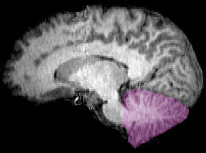
We now learn that it may have yet another function. Colleagues from Washington University School of Medicine in St. Louis report that there is a sophisticated neural computer that is buried deep in the cerebellum, that performs inertial navigation calculations to calculate our precise movement through space.
These calculations are incredibly complex and involve the vestibular system in the inner ear that provides the primary source of input to the brain about the body’s movement and orientation in space. However, the vestibular sensors in the inner ear only give us information about the position of our head position. In addition the vestibular system’s detection of head acceleration cannot distinguish between the effect of movement and that of gravitational force.
Dora Angelaki and her team based their brain studies on the predictions of a theoretical mathematical model. The model proposed that the brain could calculate inertial motion by combining two things:
1. Rotational signals from the semicircular canal in the inner ear with
2. Gravity
Based on previous research, they concentrated their search for the brain’s inertial navigation system on particular types of neurons called Purkinje cells, in a region of the cerebellum known to receive signals from the vestibular system. This region is known as the posterior cerebellar vermis, a narrow, worm-like structure between the brain’s hemispheres. It has been known for two centuries that damage to the cerebellar vermis can produce bizarre neurological problems.
In their experiments, the researchers measured the electrical activity of these Purkinje cells in monkeys as the animals’ heads were maneuvered through a precise series of rotations and accelerations. After analyzing the electrical signals measured from the Purkinje cells during these movements, the researchers concluded that the specialized Purkinje cells were, as predicted, computing earth-referenced motion from head-centered vestibular information.
This is a remarkable finding: the cells are able to make complex deductions based on very little information.
Cells in the brain, particularly cells of the Purkinje type are designed to adapt and learn. So the finding gives yet more credence to the idea that balance, coordination and orientation in space are learnable skills. That will be the topic of another research project.
But for now, the evidence suggests that anything that helps you to practice these skills will likely pay dividends as you get older.
Who’s In Charge of Your Brain?

There has been a lot of discussion in the neuroscience community about the way in which different circuits in the brain are integrated. We know that the right and left sides of the brain normally operate together, and that most of the time the three major systems of the brain: reptilian, limbic and neocortical regions work together. However, we have also speculated that one of the causes for some neurological, psychological and psychiatric illnesses might be a result of “disconnections” between different regions of the brain. The idea that some brain regions may be uncoordinated goes back more than a hundred years, but was resurrected and developed quite brilliantly in the 1960s by one of my mentors, the late Norman Geschwind, who was one of the three smartest people that I ever met.
Now there is some new research that may have important implications for neurology and psychology.
Colleagues from Washington University School of Medicine in St. Louis have found strong evidence that there is not one but two complementary commanders in charge of the brain. It is rather like having two captains in charge of the same ship.
The two captains are networks of brain regions that do not consult each other but still work toward a common purpose: the control of voluntary, goal-oriented behavior. These behaviors include a vast range of activities from walking and talking, reading and deciding on a course of action. There are yet other systems that control behaviors that are usually involuntary such as control of the pulse rate or digestion.
Last year the researchers found that there are 39 brain regions that consistently became active before the brain goes to work on a task. They did this through functional magnetic resonance imaging (fMRI) scans that follow the oxygenation of the blood. As a region of the brain is activated, blood flow increases and we can work out which brain regions are contributing to a mental task. One of the keys to our mental capacity is the ability to recruit different parts of the brain as we need them. The previous research suggested that these brain regions create task sets: plans for using the specialized talents of various brain regions to achieve a goal. Let me give you an example.
Read the word: CAT
You can do many things with that word: make a mental image of a cat, read it aloud, make rhymes or lists of words and so on. And each of those activities involves a different set of circuits in the brain. The person with good powers of attention and concentration is able to mobilize and coordinate more of his or her mental resources than the person who is less focused on a task.
In this new study the researchers used a different brain scanning technique called “resting state functional connectivity MRI.” Volunteers were asked to relax while their brains are scanned instead of working on a task.
They then used graph theory, a branch of mathematics that visually graphs relationships between pairs of objects to work out how different brain regions are coordinated. This mathematical technique is rather like the party game Six Degrees of Kevin Bacon, in which you use paired connections to go from one actor or actress to another until you’ve identified a chain of connections linking Kevin Bacon and another actor, actress or movie that wasn’t immediately obvious.
They were able to identify pairs of brain regions where blood oxygen levels rose and fell roughly in synch with each other, implying that the regions most likely work together. What they found was two separate systems. Each system had multiple connections to other regions in its own team, but they never connected to regions on the opposite team.
Having established the existence of two control networks, the next task was to work out their roles. One network – the “cinguloopercular network” – was linked to a “sustain” signal. It turns on when you start doing a task and stays constant while you do it. When you are finished it switches off.
The second is the frontoparietal network that is consistently active at the start of mental tasks and during the correction of errors.
In some senses this is not too much of a surprise. Many systems of the body have multiple control systems. I have often talked about the scores of systems involved in the control of body weight, which means that a dietetic approach directed at only one system is doomed to failure. If the body thinks that it is starving other systems jump in to slow your metabolism, reduce your activity level and make you crave certain foods.
Another good example is body temperature. Your temperature is regulated by several independent factors including your sweat glands, metabolism and activity level. If one of them goes wrong, other systems will try and compensate. When one controlling factor goes awry, others can try to compensate. The controls of your weight and body temperature are known as complex adaptive systems, and they are found not only in biology, but also in ecology, economics and atmospheric dynamics. We study them using an approach called network dynamics.
The findings may help our efforts to understand the effects of brain injury and develop new strategies to treat such injuries.
They may also help us understand problems like goal-directed behavior and human motivation.
If only one of the two systems has agreed that you need to go out and exercise, it may help us design new strategies to help you and explain why some approaches to, say, diet and exercise, are more effective than others.
Groundbreaking stuff!
Brain Training and the Paradox of Aging

For centuries, people in cultures throughout the world have believed that increasing age is associated with increasing wisdom. Not just knowledge and experience, but wisdom. Is this an old wives’ tale, or is there something to it?
There is growing evidence that as we age our brains actually become stronger. Everyone missed it, because the emphasis was on memory, concentration and thinking speed, and those may all deteriorate with age. What changes is that as we age we become more efficient at making associations rather than thinking linearly. we also recruit regions of the brain that may have lain dormant for years. At the same time, different types of mental training, including meditation, can improve many aspects of our cognitive abilities, and may also enrich and even grow appropriate regions of the brain.
Some new data from a study being conducted at Wake Forest University Baptist Medical Center in North Carolina suggests that it is possible to use a fitness program for your brain that can improve thinking skills, attention and concentration as reliably as lifting weights can increase muscle strength.
Preliminary findings from the study, which used functional magnetic resonance imaging (fMRI) to record brain activity, were presented last week at the Organization for Human Brain Mapping conference in Chicago.
As we age, we do experience changes in how we perceive the information that our senses gather from the environment. As I mentioned older adults combine information from the different senses more readily than do younger adults. This is known as sensory integration, and the down side is that it can lead to difficulties in blocking out distracting sights and sounds while still maintaining focus on important information. You are probably familiar with the “Cocktail Party Phenomenon.” You are in a loud room, but because someone is saying something interesting, you are able to block out uninteresting information. This technical term for this is cortricofugal inhibition. As we get older it gets more difficult to do the inhibiting.
The Brain Fitness in Older Adults (B-fit) study has been designed to establish whether eight hours of brain exercise can improve the ability of healthy older adults, ages 65 to 75 years, to filter out unwanted sights and sounds.
The B-fit study uses fMRI to visualize blood flow and brain activity to determine how attention training affects brain function. The training involves either a structured one-on-one mental work-out program or a group brain exercise program. During the one-on-one sessions, the volunteers were asked to ignore distracting information and tasks get harder as the eight-week training progresses. For the group sessions, participants learn new information relevant to healthy aging and are then tested on their ability to apply the new information.
All participants had an fMRI scan during a distraction task. They had to look for target words or numbers while ignoring distracting sounds. The scans showed brain activity in areas related to both sight and sound. Follow-up fMRIs showed that in the group receiving the one-on-one training, activity related to sight was increased, while activity related to sound was decreased. In addition, performance on the task was improved.
So the data do indeed suggest that attention training is a way to reduce
older adults’ susceptibility to distracting stimuli and therefore help improve
concentration.
And what about wisdom?
I have written in Healing, Meaning and Purpose that “Wisdom is the integration of understanding:” making more new associations and drawing conclusions based on a new perspective is a gift of aging. The training enables us to make sense of and communicate our conclusions.
When it comes to the brain, use it or lose!
A New Treatment for Parkinson’s Disease?

It is remarkable how often we find that a treatment for one problem may turn out to help something completely different.
Parkinson’s disease is caused by a loss of dopamine neurons in specific circuits in the brain. Dopamine has a great many actions, but I these particular circuits it is involved in the control of movement. Once it starts, the process usually continues on inexorably. The mystery has been why the neurons die in the first place.
There is a very promising article that was published online today in the journal Nature.
Investigators from the Department of Physiology in the Feinberg School of Medicine at Northwestern University in Chicago and the Department of Cellular and Molecular Pharmacology at the Chicago Medical School, Rosalind Franklin University of Medicine and Science have looked at a medicine that is usually used for treating hypertension, and found that it may protect dopaminergic neurons.
Their studies have suggested that the dopaminergic neurons involved are unusually reliant on a type of calcium channel that maintains their rhythmic activity. If the pacemaker is damaged, the neurons become more vulnerable to stressors such as neurotoxins. As we age, calcium ions begin to enter the neurons and change their behavior. They showed that mice that had been engineered to develop a progressive Parkinson’s-type disease did not become ill when their condition was treated with the calcium channel blocker israpidine. Their dopamine neurons appeared to revert back to their original, youthful form.
It is much to early to think about using isradipine for treatment. We do not, for example, know whether it works in humans and we have any idea how much of the drug would be needed. But this research does suggest a whole new line of research and possibilities for treatment.
That is exciting, though when I read about research like this I always wish that researchers did not use animals in their experiments. But since the animals have made the sacrifice to help us, we need to acknowledge their help.
Negative Ions to Help Treat Mania

Last year I talked about some of the nasty problems that can be caused by the hot dry winds that blow in many parts of the planet.
I have just seen that a paper that I was asked to look at a few months ago has just appeared in print, though according to the journal it came out in February!
The investigators used a negative ionizer to augment the treatment of 20 acutely manic people. It was a standard double-blind crossover study with raters who did not know who had received what treatment.
The negative ions did indeed produce a significant improvement in manic symptoms. We already know that atmospheric ions can have an impact on serotonin levels in some parts of the brain, and that could be the mechanism of action.
It is surprising that there has been so little research into such a simple intervention, though it is important to note that the patients were also receiving medications. But it would be valuable to see if such a simple environmental change could modulate serious mood problems before they get to the level of needing hospitalization.
Neurological Complications of Gastric Bypass Surgery

Our bodies are highly complex systems. They are not just bags of randomly assorted organs. So tinkering with one part of the system can have an impact in an entirely different part of the body. Many of us have worried for some time about the long-term consequences of gastric bypass surgery for weight loss. We know that some people who have had the surgery develop “substitution addictions.” If they had a real addiction to food before the surgery, after it they may begin to develop an addiction to something else, such as drugs or alcohol.
Now neurologists at the University of Arkansas for Medical Sciences (UAMS) in Little Rock have reported the results of a ten-year study found a link between gastric bypass surgery and several serious neurological conditions.
The study was published online May 22 in the medical journal Neurology and concludes that patients who undergo gastric bypass surgery, also known as bariatric surgery, are at risk for long-term vitamin and mineral deficiencies and as a result may develop a variety of neurological symptoms.
We know that ever more of these operations are being done every year, and so long as people are motivated they are usually successful in reducing weight. But they are not without risk, and we always have to balance that risk against the risk of being morbidly obese. This work is important because it suggests that there is an extra risk about which we previously knew very little. Many of the complications that patients experience affect the nervous system, and they are often disabling and irreversible.
More than 150 patients who came to the UAMS Neurology Clinic following gastric bypass were included in the report. In 26 of these patients a link between the surgery and their neurological condition was found.
All of the patients involved in the study had previously undergone what is known as the Roux-en-Y gastric bypass procedure in which a small stomach pouch is created by stapling part of the stomach together and bypassing part of the small bowel, resulting in reduced food intake and a decreased ability to absorb the nutrients in food. The interval between surgery and onset of neurological symptoms ranged from 4 weeks to 18 years.
The neurological problems involved many different parts of the nervous system, and the symptoms included confusion, auditory hallucinations, optic neuropathy, weakness and loss of sensation in the legs, and pain in the feet, among other conditions. None of the patients had prior neurological symptoms.
Many of the patients also experienced multiple nutritional abnormalities, especially low serum copper, vitamin B12, vitamin D, iron and calcium.
This study underlines something very important. Many heavy people are actually malnourished, because they eat the wrong things and an excess of fat in the diet can create problems in the absorption of some vitamins and minerals. In addition, surgery like this can cause a form of malabsorption.
It is therefore essential to make sure that people who have this kind of surgery have an adequate long-term intake of vitamin and mineral supplements to prevent these neurological complications. It is important for everyone to know about these potential problems and to be on the lookout for neurological symptoms.
Meditation and Learning to Pay Attention

I have talked before about the increasing difficulties that we all have with paying attention.
Over the years we have tried all sorts of things to help people to become better at focusing and controlling their attention, from games, to biofeedback devices to meditation. It is the last of those that seems to be the most effective.
We know from personal experience that paying close attention to one thing can keep us from noticing something else. If we are shown two visual signals half a second apart, we usually miss the second because we are still focused on the first one. We are consciously unaware of the second flash. It is rather like missing something when we blink our eyes, and indeed psychologists call it “attentional blink.” Many psychologists have assumed that attention is something fixed: we can only give a certain amount of attention to one thing at a time before we have to move onto something else or get a kind of brain freeze.
But we have known for some time now that sometimes people do notice the second flash of light, and with practice they can notice it all the time, suggesting that the limitation on seeing the two flashes is not entirely physical, but that it may be possible to bring it under mental control. The first style of meditation that I ever learned was Vipassana, and I well remember being astonished at how quickly my attention and focus began to improve, even when I was not meditating.
Now there is an important new study from the University of Wisconsin-Madison that suggests that attention does not have a fixed capacity, and that it can be improved by directed mental training such as meditation.
The work was done in Richard Davidson’s lab at the University of Wisconsin-Madison School of Medicine and Public Health and the Waisman Center for Brain Imaging and Behavior and published online in the journal PLoS Biology. (This is one of the growing number of open access journals, and for people not yet convinced of the value of open journals have a look at this paper, in which everything is available to you for free.)
The investigators recruited people who were interested in meditation to study whether conscious mental training can affect attention.
They examined the effects of three months of intensive training in Vipassana meditation, which focuses on reducing mental distraction and improving sensory awareness.
Volunteers were asked to look for target numbers that were mixed into a series of distracting letters and quickly flashed on a screen. As subjects performed the task, their brain activity was recorded with electrodes placed on the scalp. In some cases, two target numbers appeared in the series less than one-half second apart – close enough to fall within the typical attentional blink window.
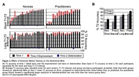
The researchers found that the three months of rigorous training in Vipassana meditation improved people’s ability to detect a second target within the half-second time window. In addition, though the ability to see the first target did not change, the mental training reduced the amount of brain activity associated with seeing the first target.
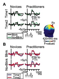
Because the subjects were not meditating during the test, their improvement suggests that prior training can cause lasting changes in how people allocate their mental resources.
As Richard Davidson says,
“Their previous practice of meditation is influencing their performance on this task. The conventional view is that attentional resources are limited. This shows that attention capabilities can be enhanced through learning.”
The finding that attention is a flexible skill opens up many possibilities. It provides further evidence that attention training is worth examining for attentional problems such as attention deficit hyperactivity disorder.
“When we raise ourselves through meditation to what unites us with the spirit, we quicken something within us that is eternal and unlimited by birth and death. Once we have experienced this eternal part in us, we can no longer doubt its existence. Meditation is thus the way to knowing and beholding the eternal, indestructible, essential center of our being.”
–Rudolf Steiner (Croatian-born Austrian Mystic, Occultist, Social Philosopher, Architect and Founder of Anthroposophy, 1861-1925)
“The moment one gives close attention to anything, even a blade of grass, it becomes a mysterious, awesome, indescribably magnificent world in itself.”
–Henry Miller (American Writer, 1891-1980)
“The path of spiritual attention is not easy, although anyone can make a beginning by trying to understand.”
— Sri Raghavan Iyer (Indian-born Prodigy, Rhodes Scholar, Academic, Philosopher, Theosophist, and, from 1965-1986, Professor of Political Science at the University of California, Santa Barbara, and Father of Pico Iyer, 1930-1995)
Vyvanse
There is no doubt that we need more options for the treatment of attention-deficit/hyperactivity disorder (ADHD), and though it would be nice to be able to treat everyone with a diet or some other approach, for many it is just not feasible.
Of the current medicines, most do not last quite long enough, and occasionally may need to be taken more than once a day. The trouble is that taking these medicines intermittently, or only having it in the circulation for a short time, plays havoc with the dopamine receptors in the brain.
One of the big problems with the current group of stimulant medicines is that they are amphetamines, and have enormous abuse potential. They are sold and re-sold and taken by mouth, inhaled, smoked and injected. In some parts of the United States abuse of ADHD medicines is the number one form of substance abuse.
So it would be a huge advantage to have long-lasting medicines with a much-reduced risk of abuse.
Vyvanse is an extremely interesting new treatment for ADHD that has just been approved by the FDA and should be available in the United States in the very near future. It is manufactured by Shire Pharmaceuticals that is getting a reputation as the company with the biggest interest in ADHD.
During development, Vyvanse was known as NRP104. Its main ingredient is lisdexamfetamine dimesylate. Vyvanse is unique in another way too: it is a prodrug or ‘conditionally bioreversible derivative’ of amphetamine, the main ingredient in Adderall and Adderall XR. This is a key point: unlike other stimulants it is conjugated to an amino acid, and has to go through the stomach and be digested before it can become active. Therefore there is a much reduced risk that it will be abused, since it cannot be snorted or injected like other ADHD medicines.
There was a great deal of new data presented at the In research presented this week at the 2007 Annual Meeting of the American Psychiatric Association in San Diego, California. The bottom line seems to be that the medicine is both effect and well tolerated. It is going to be a genuinely useful addition to the treatments that we can offer people with ADHD.


