Low Testosterone May Shrink Your… Brain

It is almost forty years since Fernando Nottebohm first began to describe some of the dynamic changes that occur in the brains of songbirds as the seasons change. Every year some regions of the brain grow in response to changes in ambient light levels and others regress. There are marked seasonal changes in the brains of fish, reptiles, amphibians, birds and even some mammals such as gerbils, mice and perhaps even in humans. But the magnitude of the changes in birds far outweighs any other species. It is hoped that that understanding the mechanism controlling that change may help us to develop treatments for age-related degenerative diseases of the brain such as Parkinson’s and dementia.
Researchers from the University of Washington and the University of California, Berkeley, have published some interesting new data in the Proceedings of the National Academy of Sciences. They report a striking shrinkage in the size of the brain regions that control singing behavior of Gambel’s white-crowned sparrows. This transformation is triggered by the withdrawal of testosterone and can be seen within 12 hours. The study is the first to report such rapid regression of brain nuclei caused by the withdrawal of a hormone and a change in daylight conditions in adult animals.
The research protocol was designed to mimic the natural seasonal changes that occur in the brains of the sparrows. Their song-control regions expand in the spring and summer leading up to the breeding season, as they use songs to establish territories and attract mates in Alaska. Later in the summer, as the birds get ready to migrate back to California, the same brain regions shrink.
To better understand what happens in the sparrows’ brain, the researchers received federal and state permits to capture 25 of the migrating male birds in Eastern Washington. They then housed the birds for 12 weeks before exposing them to 20 days of long-day conditions comparable to the natural lighting the sparrows would experience in Alaska during the breeding season. The birds were also implanted with testosterone.
At the end of 20 days, six of the birds were euthanized and the remaining 19 were castrated and testosterone implants were removed so there would not be any circulating testosterone in their systems. After 12 hours five more birds were euthanized and the remainder were euthanized at 2, 4, 7 and 20 days.
The researchers found that the size of the high vocal center (HVC) region decreased 22 percent within 12 hours after the withdrawal of testosterone and that the number of neurons in this song-control region fell by 26 percent after four days. In addition, the size of two other song-control regions called Area X and the RA significantly regressed after 7 and 20 days.
Much as I dislike animal experiments of any kind, it is invaluable to have an animal model system in which we can observe predictable neurodegeneration. As men age, circulating levels of testosterone decrease, and other research has shown that this decline may contribute to cognitive impairment.
This is an important new approach to understanding the interplay between nerve cell degeneration and hormones.
Brain-Derived Growth Factors and Bipolar Disorder
As we are learning more about the plasticity of the brain, and the way in which new neurons can continue to grow throughout life, there is a great deal of interest in factors that stimulate the growth or development of neurons. This is particularly important in condition like schizophrenia and bipolar in which cognitive decline may occur.
Researchers from Portugal presented some interesting new data (NR68) on Monday at the 2007 Annual Meeting of the American Psychiatric Association in San Diego, California.
Genetic and pharmacological studies have suggested that brain-derived neurotrophic factor (BDNF), one of the most common neurotrophic factors in the brain, may be associated with the pathophysiology of bipolar disorder. The data has been a bit of a mixed bag: previous studies have suggested that BDNF may be associated with either a worse or better neurocognitive outcome in tests of frontal lobe function.
The researchers examined 28 people with bipolar disorder whose mood was currently normal: i.e. they were euthymic, and they compared them with 25 healthy volunteers. They measured BDNF levels and performed a battery of neuropsychological tests.
The people with bipolar disorder had clear evidence of problems with attention and executive function even when their mood was normal. However it does not seem to have much to do with BDNF: the levels were the same in both patients and healthy volunteers. There was an association between BDNF levels and memory in people with bipolar disorder. This makes sense: BDNF has been implicated in the formation of memory traces in the brain.
What this means is that problems of attention and executive function are likely to be trait-markers of bipolar disorder, while BDNF levels may be a state-related biological marker.
It is interesting how the wheel keeps turning: the person who first differentiated dementia praecox (schizophrenia) from manic depression (bipolar disorder) was Emil Kraepelin. He originally said that people with dementia praecox became worse over time, because of progressive cognitive decline, while people with manic depression did not. When he passed away in 1926 he was working on a whole re-visioning of mental illness, saying that the two could not be so clearly differentiated, because cognitive decline could occur in either illness.
Many pharmaceutical companies are looking into the possibility of improving cognition in major mental illnesses, but there are also a great many non-pharmacological interventions that can be done to help cognition in people with major mental illness.
Lithium and Brain Growth

There’s an interesting story about the way that lithium was found to be helpful to some people with bipolar disorder. It was thought that there was a link between bipolar disorder and gout, and gout is caused by an accumulation of uric acid in some joints. It just so happens that lithium is very good at getting guinea pigs to pass any excess uric acid in their urine. So in 1949 a medical scientist in Australia called John Cade tried lithium in bipolar disorder. It worked, but not because of any effect on uric acid: the lithium/uric acid connection only works in guinea pigs. But now, all these years later, it turns out that there may actually be a link between bipolar disorder and uric acid after all: uric acid levels often rise in people who are insulin resistant, and insulin resistance is a common feature of bipolar disorder.
The last few years have seen the growth of an impressive body of evidence about the effects of lithium on the brain. Lithium modulates the way in which many neurotransmitters act in the brain. That raised interesting questions, because many of the chemical transmitters do not only send messages between neurons, they may also stimulate the growth of cell processes and perhaps neurons themselves. Then it was discovered that lithium protects neurons by increasing the levels of a neuroprotective protein called Bcl-2. Then it was found to help stimulate the production of new neurons (neurogenesis) in the hippocampus.
A major breakthrough came in 2000, with the demonstration that lithium increases the amount of gray matter in the human brain, probably by stimulating the production of a growth promoter called brain-derived neurotrophic factor. Gray matter is composed primarily of neurons, so this suggested that lithium was either stimulating the growth of new neurons, or causing the migration of neural stem cells, or alternatively somehow preventing the death of neurons.
Within less than two years it was confirmed that treatment with lithium increased the amount of gray matter in the brains of people with bipolar disorder.
The findings remained controversial for two reasons: first was the seeming impossibility of stimulating the production of new gray matter. That is no longer such a problem: there is more and more evidence that the adult brain can indeed produce new neurons.
The second problem was technology: the measurements were open to
different interpretations.
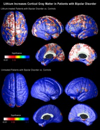
Now neuroscientists at UCLA, using brain scans collected by collaborators at the University of Pittsburgh School of Medicine have shown that lithium does indeed increase the amount of gray matter in the brains of patients with bipolar disorder. But what is far more important is that the effects are seen in precise regions of the brain that are thought to be implicated in bipolar disorder.
The research is published in the July issue of the journal Biological Psychiatry.
Carrie Bearden, a clinical neuropsychologist and assistant professor of psychiatry at UCLA, and Paul Thompson, who is associate professor of neurology at the UCLA Laboratory of NeuroImaging, used a novel method of three-dimensional magnetic resonance imaging (MRI) to map the entire surface of the brain in people diagnosed with bipolar disorder. Everyone interested in the brain has been amazed by the quality of the images coming from the UCLA lab over the last six or seven years. (Graphics from the study, including the one above, are available online here.)
When the researchers compared the brains of bipolar patients on lithium with those of people without the disorder and those of bipolar patients not on lithium, they found that the volume of gray matter in the brains of those on lithium was as much as 15 percent higher in the cingulate and paralimbic regions of the brain, that are critical for attention, motivation and emotional control. There was no change in the amount of white matter, telling us that these effects are unlikely to be either artifacts or swelling of the brain.
These are extraordinary findings that may help explain how lithium works. If lithium increases the amount of gray matter in specific regions of the brain, this may suggest that existing gray matter in these regions may be underused or dysfunctional in people with bipolar disorder.
Conventional MRI studies in bipolar disorder measured the total volume of the brain, but these new imaging techniques employ high-resolution MRI and cortical pattern-matching methods to map gray matter differences that are so precise that they can pick up subtle differences in brain structure and this is the first time that researchers have been able to look at specific regions of the brain in people with bipolar disorder, and examine the effects of treatment.
The researchers at UCLA analyzed MRI scans of 28 adults with bipolar disorder, 20 of whom were lithium-treated, and 28 healthy control subjects collected by collaborators at the University of Pittsburgh. The analysis involved detailed spatial analyses of the distribution of gray matter by measuring local volumes of gray matter at thousands of locations in the brain.
This is superb research, and one of the signs of good research is that it raises yet more questions:
- We do not know whether these effects on gray matter persist once the medicine is stopped.
- Neither do we know whether the same effect might occur in other illnesses in which there is loss of gray matter. It may just be that lithium corrects an abnormality that is specific to bipolar disorder. That being said, there is recent evidence that lithium may be neuroprotective in a mouse model of Alzheimer’s disease.
- We also do not know if these brain changes correlate with clinical improvement.
- Lithium can have a lot of side effects, so we shall also need to factor that into the equation.
- We also need to know whether other medicines – or any other kind of therapy – might produce similar effects: there are already several medicines that have been shown to do something similar in some animals.
It’s still a bit too early to suggest putting lithium in the water supply…
A Major Breakthrough in Neurogenesis Research

I have written a lot about adult neurogenesis, because it is so incredibly important.
When most of us were in school were taught that the brain is a static structure. That you are born with a certain number of neurons, that constitutes your neural “stash.” And after the age of five you would lose a hundred thousand neurons a day, and if you drank too many martinis it would be two hundred thousand a day. “Facts” and figures repeated from one textbook to another for fifty years. And quite wrong.
Ten years ago it was discovered that there are neural stem cells in the hippocampi of adult rodents. A study published in 1998 provided some evidence that neurogenesis takes place in the adult human hippocampus, but the idea that neurogenesis takes place in adult humans has remained contentious.
Now, an advance online publication on the website of the journal Science provides evidence that neurogenesis may also occur in the olfactory bulb of the human brain.
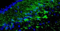
The whole article, including the pictures is available here. The picture at the top of this article shows new cells appearing in the olfactor bulb.
The investigators examined the rostral migratory stream (RMS) which is the main pathway by which newly born cells from the subventricular zone (SVZ) reach the olfactory bulb in rodents. The researchers discovered that there is a human RMS containing neuroblasts, a.k.a. neural stem cells.

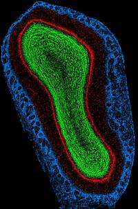
(This is a rat olfactory bulb)
Maurice Curtis and his colleagues examined the brains of cancer patients who had recently died, after having been previously injected with bromodeoxyuridine (BrdU), a chemical that is incorporated into newly synthesized DNA. Cell biologists and oncologists have used the method for years to visualize and monitor the growth of tumors. The researchers found BrdU-positive cells in the olfactory bulbs of the patients’ brains, indicating that they contained newly-generated neurons.
The team then used antibody staining to show that the neuroblasts begin to differentiate into olfactory neurons while migrating through the rostral migratory stream. Upon arriving at the bulb, the cells continued to differentiate, forming mature olfactory neurons.
The patients whose brains were examined were between 38-70 years of age, so the findings show that neurogenesis may occur throughout the duration of the human lifespan. The function of these newly generated cells is unclear, but they may be involved in recognizing and remembering new smells in the later years of life.
The researchers also have preliminary unpublished data that the stem cells not only migrate to the olfactory bulb, but also leave the RMS and migrate into the basal ganglia and cerebral cortex. This is significant, because parts of the basal ganglia degenerate in movement disorders such as Parkinson’s Disease, and specific regions of the cortex degenerate in Alzheimer’s. The possibility that stem cells enter these regions from the RMS could therefore provide a means for developing new treatments for neurodegenerative diseases.
So although at the moment new neurons are only known to be created in a small number of ancient structures in the brain, but that may well change, since the cells are marching over there from regions around the ventricles. After all, why would Nature, the Universe or God have left neural stem cells sitting there unless it was for a good reason? And if adult neurogenesis can be demonstrated in other parts of the brain, then what? An end to the stem cell debates? If the stem cells are already present in the brain, the effort can be directed toward activating and directing them. Could we continue generating new neurons to stave off the dementing illnesses, or is that just science fiction?
The answer is that science fiction is fast becoming science fact. This research will need to be replicated, but I know of several other labs that have already accumulated some similar data to this new paper in Science.
Learning to Tango

As a youngster I was introduced to most of the social graces: piano lessons – which sadly I hated – shooting, horse riding, fencing and ballroom dancing. And I just loved to tango!
Now I learn that it is, perhaps, something that more people should try.
Colleagues from the University of Turin used functional magnetic resonance imaging to study the hypothesis that simply focusing attention on walking could modify the activity of specific regions of the brain. But instead of using regular walking they taught people to tango. (The whole paper is available online as a free download.)
If you think about it for a moment, regular walking is an automatic process that you learned as a small child. It involves a series of structure in the deep – subcortical – regions of the brain and the spinal cord. The researchers got their experimental subjects to learn the dance by a combination of physical and mental practice. They were asked to perform the basic steps of the tango followed by imagining that – without moving – they were doing the steps.
The experiment showed that the training was associated with an expansion of regions of the brain involved in imagining movement. After the training the visual imagery gradually gave way to motor and kinesthetic ones.
This makes good sense: we normally begin by visualizing and then over time a motor skill like this becomes automatic. I’ve known good dancers who could flip from one dance to another without a moment’s thought.
This new finding suggests that rehabilitation of people who have lost the ability to walk should involve not just the motor act of walking, but also visualization.
But notice that the two processes – visualization and motor learning are separate. This is another nail in the coffin of the popular idea that your brain cannot tell the difference between a real and an imagined activity.
It also provides further evidence that both mental and physical activities can expand and activate regions of the brain.
And what a fun way to do it!
“The truest expression of a people is in its dance and in its music. Bodies never lie.”
–Agnes de Mille (American Choreographer, 1905-1993)
Healing the Broken Brain

I recently reviewed a fascinating book on adult neurogenesis: the creation of new neurons; something that was thought to be impossible until very recently. It is still thought to occur in certain specific regions of the brain, but even that may be changing.
The field is moving forward very rapidly and is important to every one of us, which is the reason for writing so much about the topic.
There is an exceptionally interesting article in the current issue of the journal Neuron. Researchers from Lund in Sweden have shown that cells generated from stem cells in an adult, diseased and damaged brain function as normal nerve cells. Not only do the new cells function like proper neurons, they also try to make connections with other neurons, indicating that they are trying to repair, or compensate for diseased or damaged parts of the brain.
This work was done in rats and is in its infancy, but it part of a global effort to learn more about how new neurons are formed, how they function and whether it is possible to help the brain heal itself after a disease or injury.
I am going to go out on a limb and say that it is possible. In Healing, Meaning and Purpose I described an individual with a severe neurological problem that responded to a novel treatment method using the subtle systems of the body. One of the goals of Integrated Medicine is to establish how best to use such methods in combination with conventional medicine not just to treat someone, but to initiate healing.
“A therapist doesn’t heal, he lets healing be.”
–A Course in Miracles (Book of Spiritual Principles Scribed by Dr. Helen Schucman between 1965 and 1975, and First Published in 1976)
Links, Citations and References
Following my recent article about mystical experiences, I had a charming note which included this question:
"Could you kindly give a reference or link to the above quote from Ramana Maharshi?"
This was my response:
"I am traveling at the moment, so I don’t have access to my library.
Happily I remember exactly where the quote came from. It was from someone who taught me a great deal – the late Paul Brunton. I think that Paul probably did more to introduce Ramana Maharshi to the West than anyone else, and he was also a close friend of Aurobindo. He once commented that it was rather convenient that two of the greatest sages of the 20th century only lived 65 miles apart!
He also mentions Ramana’s comment in Chapter 9, paragraph 23 of Volume 16 of The Notebooks of Paul Brunton.
Here’s the exact quote:
"That these differences of view exist even among illumined mystics is a striking but rarely studied fact. Why did Ramana Maharshi poke gentle fun at Aurobindo’s doctrine of spiritual planes?"I do not know off hand whether there are any websites that specifically discuss their – respectful – differences of opinion.
If that does not give you what you need, please do let me know: I shall be home in a couple of days and I’d be happy to do a bit more digging in my library: I have over 12,000 volumes, and there are quite a lot of lesser known titles in there. Since I raised it, I shall also have a look online when I get home.”
The reason for re-printing this correspondence is this. Regular readers will know that my blog entries are festooned with links, citations and references. That is very deliberate. This blog is a bit different from most of the other medical ones out there in that I try to ensure that you can check everything that I say.
The other day I had the privilege of speaking to the Rotary Club of New York and mentioned that I had written something about five strategies that can dramatically reduce your risk of developing Alzheimer’s disease as well as discussing some of the evidence concerning nutrition and neurogenesis: the production of new neurons. I was asked what they were, and I recommended that people asking the question should check out the article. The reason was simply this: I wanted them to see the evidence for themselves.
I think that we have all had enough of people simply expressing opinions.
Now is the time for people who are writing on line – or anywhere else for that matter – to provide data to support what they are saying.
If you need further reading material on any of the topics that I write about, just let me know: I have access to a great many sources that are not always easy to get at.
Neural Stem Cells
Stem cell research presents us with one of the most difficult moral and ethical dilemmas. Most of us have our opinions about the whole matter. But there is now a very real possibility that the scientific piece may be resolved in a different way altogether.
A recent study in the Proceedings of the National Academy of Sciences has confirmed that in adult mammals, the regions of the brain below the ventricles – the fluid-filled spaces – of the brain, harbor neural stem cells. The evidence for their existence has been building over the last five years. But this latest report is of great interest. The scientists were able to show that a soluble carbohydrate-binding protein named galectin-1 promotes the proliferation of these neural stem cells in the adult brain. These neural stem cells are highly active in the forebrains of mice.
We have yet to discover their role in adult human brains. But it would seem a safe bet that they will if anything be more active in humans, since we are endowed with incredible neural plasticity that is way beyond anything seen in most other species. And we would therefore expect to see more potential for neurogenesis in the human brain. It is valuable to place this new finding in the context of experimental work indicating that some medicines may stimulate adult neurogenesis.
“I’ve found that the chief difficulty for most people was to realize that they had really heard new things: that is things that they had never heard before. They kept translating what they heard into their habitual language. They had ceased to hope and believe there might be anything new.”
–Peter Demianovich Ouspensky (Russian Philosopher, 1878-1947)
Technorati tags: Stem cells Neurogenesis Adult brain
Growing Your Brain
Several years ago we learned that London taxi drivers grew a small part of their hippocampus while learning “The Knowledge,” a detailed map of the streets of the city. Unlike most American cities, London is like a rabbit warren that has been growing organically for two thousand years. Despite the Great Fire of 1666, and the best efforts of Hitler’s Luftwaffe, the basic illogical structure has just become more complex over the centuries. The hippocampus is intimately involved in memory, navigation and in constructing a map of your surroundings. It has also been established that professional musicians and people who use Braille, all have increases in the size and complexity of a specific region of the brain.
We now learn from researchers in Germany that medical students show an increase in the amount of grey matter in the posterior part of the hippocampus and also in regions of the parietal lobes while studying for their exams. It is calculated that a medical student learns around 6,000 new words during his or her training, so it is no surprise that the brain changes to accommodate all this new information.
This new finding just adds to our knowledge about the plasticity of the brain. Even if you do not have to learn 6,000 new words and innumerable new concepts and practical techniques, it is worth knowing that learning any new knowledge or skills will likely grow specific regions of your brain.
So make a resolution to learn at least one new thing each day, and to work on developing a new skill. It doesn’t matter whether it is learning to knit or how to play chess. Each will likely help you.
Later this year we will be bringing out a book that teaches brain stretching skills based on the latest advances in brain science.
Watch this space!
Technorati tags: Hippocampus Parietal lobe Learning Medical students Neurogenesis
Psychotropic Medicines and Neurogenesis
One of the genuine breakthroughs of the last few years has been the understanding that the brain is not a static structure, but is instead an organ that grows, refashions and repairs itself to a remarkable degree.
You may be interested in a brief review article looking at the effects of some of the medicines that appear able to stimulate neurogenesis in the adult brain.
It is no longer far-fetched science fiction to say that we are likely soon to be able to regenerate parts of the brain and spinal cord that have been damaged by disease or trauma. And in the meantime, we have an array of stratgeies that you can adopt to keep your brain active throughout life, while dramatically reducing your risk of developing dementia.
Technorati tags: Neurogenesis Antipsychotics Antidepressants Brain repair


